Sports Medicine Specialists for Women & Girls in Bay Area & West Coast
Welcome to The UCSF Women’s Sports Medicine Center
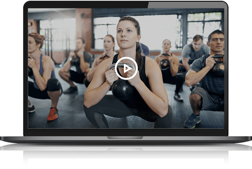
UCSF ranks among the nation's best. We provide nationally renowned sports medicine care for women, by women. Our mission is to enhance musculoskeletal health in female athletes through a multidisciplinary approach that optimizes performance and minimizes injury risk.
Areas of Expertise
Our specialties include treating ACL tears, shoulder instability, rotator cuff tears, elbow injuries, hip impingement, labral tears, overuse injuries, acute injuries, patellofemoral pain, stress fractures, and dislocations.
Board-Certified & Fellowship-Trained Sports Medicine Specialists
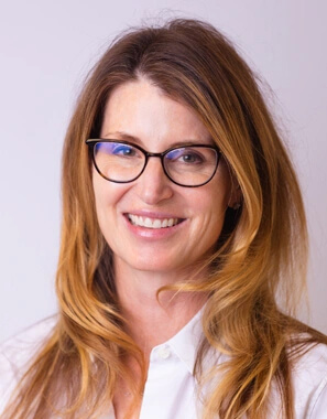
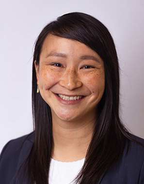
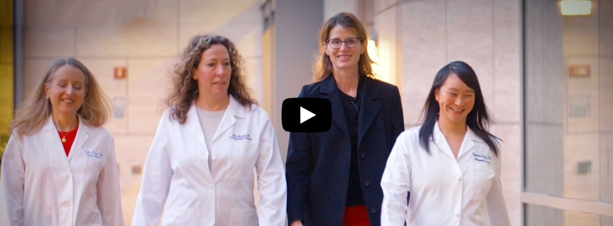
Meet Our Team
UCSF Women’s Sports Medicine Center has a multidisciplinary team of orthopedic surgeons, primary care sports medicine physicians, sports dietitians, and physical therapists, working together for your benefit.
Profiles


Orthopedic Care for Women, by Women
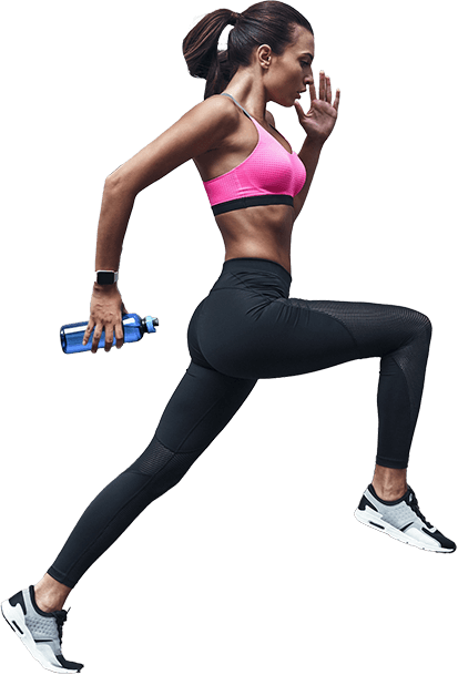
Comprehensive Support for Female Athletes
Our Multidisciplinary team offers comprehensive treatment to female athletes across the lifespan. We are Focused on the unique needs of women, from bone health to injury prevention and recovery.

Structured treatment plans designed to restore strength, flexibility, and function after injury or surgery. Tailored to each patient’s condition and recovery goals, these protocols guide safe progression through exercises and techniques that reduce pain, improve mobility, and support long-term musculoskeletal health while injury prevention.

A Compassionate, Human Experience.
Latest Updates & Insights
-
EventsEmpowering Young Athletes: Injury Prevention and Nutrition Clinic for Seals Girls’ Soccer by Physical Therapist Erin Finestone and Sports Dietitian Lindsay Orbeta
-
EventsUCSF Hosts The Perry Initiative Medical Student Outreach Program to Empower Future Female Orthopedic Surgeons
-
In the MediaThe ‘single most important behavior’ for women in perimenopause and menopause
-
In the MediaLindsay Orbeta appears on Celutions Education podcast episode, ‘Carb for Runners: Go Low or Aim High’
-
PublicationsPregnancy-Related Hip and Pelvis Musculoskeletal Conditions, Risk Factors, and Prevention
-
PublicationsAtopic Dermatitis and Tobacco Smoke Exposure During Childhood and Adolescence